Reflecting the inside out: a 3D approach to jaw surgery
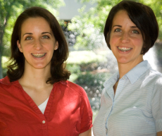 |
Rebecca Squires and Amanda Bartelme are mirror-image identical twins.
They appear to be exact reflections when facing each other.
Beyond looking alike, the sisters, 32, also share a misalignment of the jaws and severe underbites that have become more troublesome as they’ve gotten older. Five years of braces failed to fix the misalignment, and they became used to jaw pain, the inability to eat certain foods, and some difficulty enunciating.
When the twins both ended up in the Atlanta area after college, they decided to look into surgical options for correcting the misalignment. Their search led them to Emory oral surgeon Steven Roser.
The timing felt right, says Bartelme, who recently married: “We wanted to take care of things before we started having children and also while we were relatively younger and able to heal faster.”
After a clinical examination, Roser (himself a father of twins) told them that he could, indeed, perform corrective surgery with an excellent likelihood of fixing their underbite, which would then allow their orthodontist to successfully align their teeth. He also decided that the twins’ case would be ideal for a new surgical approach using 3D virtual imaging software. Used in less than 5% of jaw surgeries, the virtual software allows surgeons to do more accurate and detailed planning before procedures to create 3D models of the face based on CT scans.
The conventional method for oral and maxillofacial surgery requires taking numerous measurements of the jaw area and casting molds of the upper and lower teeth. The molds are positioned, repositioned, and measured, allowing the surgeon to calculate needed measurements. Essentially, the doctor runs through the surgery with the models before entering the operating room.
With the new technique, an optical scan of the teeth is merged with a CT scan of the head. The surgeon and biomedical technician view the images online and together use the software to perform surgery that allows them to accurately predict the surgical skeletal movements to correct jaw alignment. The computerized method allowed Roser to create a 3D simulation.
The twins’ only request was that whatever was done to one of them be done to the other. “It was a pretty daunting surgery,” Squires says. “This was something we were going to do together or not at all.”
During more than five hours of surgery each—all from inside the mouth—the twins’ upper and lower jaws were cut and realigned, plates were inserted, and their jaws were wired shut for healing. The only downside of recovery, they say, was subsisting on a liquid diet for 25 days.
While they still need to wear braces for another six months to a year, the twins are happy with the results of the surgery.“We’re more confident in our appearance, can speak more clearly, and overall have the peace of mind that we’re preventing future issues in taking care of ourselves now,” Bartelme says.
Another benefit of the virtual surgery planning, says Roser, is that it allows for more accurate monitoring of the results. “Better planning,” he says, “leads to better outcome.”
The twins are pleased that they continue to look alike. “We’re happy still being identical,” says Bartelme. —Mary Loftus