News Release: Research, School of Medicine
Jul. 21, 2009
Nanoparticles Could Improve Breast Cancer Diagnosis Via MRI
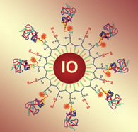 Diagram showing "nanoparticle" in question. Download larger version of graphic.
Emory researchers have developed tools for improving the diagnosis of breast cancer by attaching onto iron oxide "nanoparticles" a molecule that binds specifically to breast cancer cells.
The iron makes the particles clearly visible under magnetic resonance imaging (MRI), and thus could enhance breast MRI, a widely available diagnostic technique. At clinical field strength, tumors with specific disease signatures, implanted in mice, can be detected by MRI with the nanoparticles.
The nanoparticles' properties are described in the July 15 issue of the journal Clinical Cancer Research.
The research was a collaboration between Lily Yang, MD, associate professor of surgery at Emory University School of Medicine, Hui Mao, PhD, associate professor of radiology at Emory, and Shuming Nie, PhD, a professor in the Wallace H. Coulter Department of Biomedical Engineering at Georgia Tech and Emory University.
Recently the American Cancer Society recommended breast MRI as a screening approach, in addition to mammography, for early detection in women at high risk. Breast MRI is useful because of the high resolution and ability to differentiate tissue types, Mao says.
"Conventional breast MRI is very sensitive, but it can lack specificity, which leads to a high number of false positive findings," he says. "A MRI probe that could recognize a breast cancer based on its biomarkers would be very helpful to improve breast cancer diagnosis."
The nanoparticles have an iron oxide core that is 10 nanometers in diameter, with a polymer coating. The surfaces of the particles have attached a fragment of a natural protein found in humans: urokinase plasminogen activator (uPA), which binds to its receptor (uPAR) on several types of cancer cells. This molecule allows the particles to discriminate between breast cancer cells and healthy cells.
In mice implanted with human tumors, particles coated with a fragment of uPA are taken up by breast cancer cells and also by the liver, the researchers found. The nanoparticles stay in the blood for several hours. Uptake of the nanoparticle probes by the tumor is not uniform. The areas that display the greatest contrast with uPA-conjugated iron oxide nanoparticles are on the outer layer of the tumor, rich with blood vessels.
The technology needs to be refined and validated in animal models before it is ready to test in patients, Mao says. Women with increased risk of breast cancer, such as those with inherited cancer risk factors, could potentially benefit.
"We believe this is an important step toward developing cancer-specific breast MRI that could allow early detection and treatment monitoring," he says.
The research was supported by grants from the National Cancer Institute for cancer nanotechnology and imaging.
Reference:
Yang, L. et al Receptor-Targeted Nanoparticles for In vivo Imaging of Breast Cancer. Clin. Can. Res. 15, 4722-4732 (July 15, 2009)
###
The Robert W. Woodruff Health Sciences Center of Emory University is an academic health science and service center focused on missions of teaching, research, health care and public service. Its components include schools of medicine, nursing, and public health; Yerkes National Primate Research Center; the Emory Winship Cancer Institute; and Emory Healthcare, the largest, most comprehensive health system in Georgia. The Woodruff Health Sciences Center has a $2.3 billion budget, 17,000 employees, 2,300 full-time and 1,900 affiliated faculty, 4,300 students and trainees, and a $4.9 billion economic impact on metro Atlanta.
Learn more about Emory’s health sciences:
Blog: http://emoryhealthblog.com
Twitter: @emoryhealthsci
Web: http://emoryhealthsciences.org